Endoscopic Exam for Cancer
How is cancer diagnosed?
There is no single test that can accurately diagnose cancer. It usually requires a thorough history and physical exam along with diagnostic testing. Many tests are needed to determine whether a person has cancer, or if another condition (such as an infection) is mimicking the symptoms of cancer. Effective diagnostic testing is used to confirm or eliminate the presence of disease, monitor the disease process, and plan for and evaluate the effectiveness of treatment. In some cases, it is necessary to repeat testing when your condition has changed, if a sample collected was not of good quality, or an abnormal test result needs to be confirmed. Diagnostic procedures for cancer may include imaging, lab tests (including tests for tumor markers), tumor biopsy, endoscopic examination, surgery, or genetic testing.
What are the different types of endoscopic exams?
An endoscope is a small, flexible tube with a light and a lens or tiny video camera on the end used to look into the esophagus, stomach, duodenum, colon, rectum, or other organs. It can also be used to take tissue from the body for testing or to take color photographs of the inside of the body. Several types of endoscopes are described below:
-
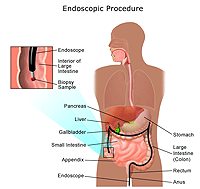
Colonoscopy. Colonoscopy is a procedure that allows the doctor to view the entire length of the large intestine (colon), and can often help identify abnormal growths, inflamed tissue, ulcers, and bleeding. It involves inserting a colonoscope, a long, flexible, lighted tube, in through the rectum up into the colon. The colonoscope allows the doctor to see the lining of the colon, remove tissue for further examination, and possibly treat some problems that are discovered.
Important information! Please call your insurance company before your colonoscopy. You will want to know how much you and your insurance will pay. The cost depends on:
 |
| Click Image to Enlarge |
-
Endoscopic retrograde cholangiopancreatography (ERCP). ERCP is a procedure that allows the doctor to diagnose and treat problems in the liver, gallbladder, bile ducts, and pancreas. The procedure combines X-ray and the use of an endoscope, which is a long, flexible, lighted tube. The scope is guided through the person's mouth and throat, then through the esophagus, stomach, and duodenum (the first part of the small intestine). The doctor can examine the inside of these organs and detect any abnormalities. A tube is then passed through the scope and a dye is injected, which will allow the internal organs to appear on an X-ray.
-
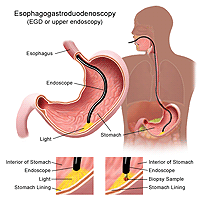
Esophagogastroduodenoscopy (also called EGD or upper endoscopy). An EGD (upper endoscopy) is a procedure that allows the doctor to examine the inside of the esophagus, stomach, and duodenum. A thin, flexible, lighted tube, called an endoscope, is guided into the mouth and throat, then into the esophagus, stomach, and duodenum. The endoscope allows the doctor to view the inside of this area of the body, as well as to insert instruments through a scope for the removal of a sample of tissue for biopsy (if necessary).
 |
| Click Image to Enlarge |
-
Sigmoidoscopy. A sigmoidoscopy is a diagnostic procedure that allows the doctor to examine the inside of a portion of the large intestine, and can be helpful in identifying the causes of diarrhea, abdominal pain, constipation, abnormal growths, and bleeding. A short, flexible, lighted tube, called a sigmoidoscope, is inserted into the intestine through the rectum. The scope blows air into the intestine to inflate it and make viewing the inside easier.
-
Bronchoscopy. A bronchoscopy is a diagnostic procedure that allows the doctor to examine the inside of the trachea (windpipe) and bronchi (large airways leading into the lungs). A short, flexible, lighted tube, called a bronchoscope, is inserted through the mouth or nose. Samples of tissue may be removed through the bronchoscope for examination under a microscope in the lab.
-
Cystoscopy. An examination in which a cystoscope, a flexible tube and viewing device, is inserted through the urethra to examine the bladder and urinary tract for structural abnormalities or obstructions, such as tumors or stones. Samples of the bladder tissue may be removed through the cystoscope for examination under a microscope in the lab.